| Subarachnoid space | |
|---|---|
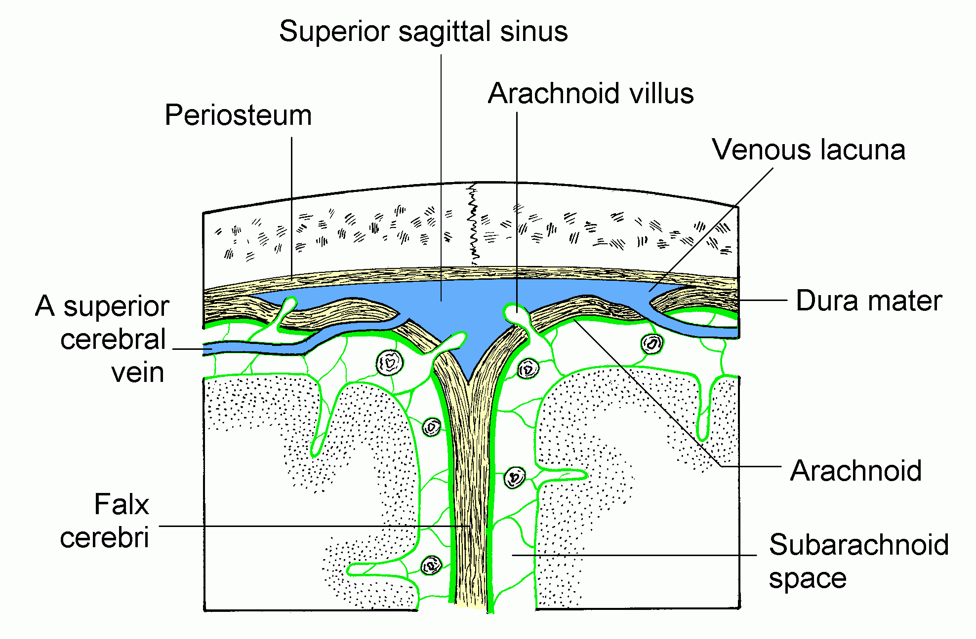
| Diagrammatic representation of a section across the top of the skull, showing the membranes of the brain, etc. ("Subarachnoid cavity" visible at left.) | |
| Diagrammatic transverse section of the medulla spinalis and its membranes. (Subarachnoid cavity colored blue.) | |
| Latin | spatium subarachnoideum, cavum subarachnoideale |
| Gray's | subject #193 876 |
| MeSH | Subarachnoid+Space |
In the central nervous system, the subarachnoid cavity (subarachnoid space) is the interval between the arachnoid membrane and pia mater.
It is occupied by spongy tissue consisting of trabeculae (delicate connective tissue filaments that extend from the arachnoid mater and blend into the pia mater) and intercommunicating channels in which the cerebrospinal fluid is contained.
This cavity is small on the surface of the hemispheres of the brain. On the summit of each gyrus the pia mater and the arachnoid are in close contact, but in the sulci between the gyri, triangular spaces are left, in which the subarachnoid trabecular tissue is found. Whilst the pia mater closely follows the surface of the brain and dips into the sulci, the arachnoid bridges across them from gyrus to gyrus.
At certain parts of the base of the brain, the arachnoid is separated from the pia mater by wide intervals, which communicate freely with each other and are named subarachnoid cisternæ; in these the subarachnoid tissue is less abundant. The subarachnoid space is the location of the interface between the vascular tissue and the cerebrospinal fluid and is active in the blood brain barrier.
The arachnoid mater continues down the spinal cord too, and the subarachnoid layer with it. It serves a similar function in the spinal cord as it does in the brain.
This article was originally based on an entry from a public domain edition of Gray's Anatomy. As such, some of the information contained within it may be outdated.
|
||||||||||||||||||||||||